
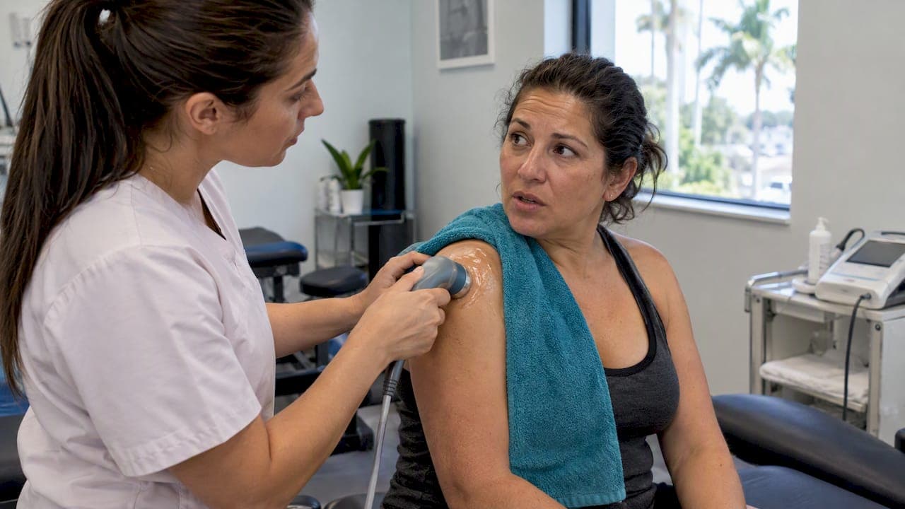
Ultrasound therapy should be avoided over the eyes, a pregnant uterus, the reproductive organs, any known malignancy, active infection, recent hemorrhage, a deep vein thrombosis, or an area with a pacemaker or other active implant. If any of these apply to you, the safe move is to pause and let a clinician clear the area first, since treating through them can worsen the underlying problem rather than relieve it.
TL;DR:
- Ultrasound therapy should be avoided over the eyes, pregnant uterus, reproductive organs, known malignancies, infections, recent hemorrhages, deep vein thrombosis, and areas with pacemakers or other implants.
- Use of ultrasound near sensitive areas requires cautious dose adjustments and continuous movement of the transducer to minimize thermal or mechanical injury.
- Clinicians must monitor thermal and mechanical safety indices, keep settings at the lowest effective levels, and document all parameters to prevent adverse effects.
- Contraindications include areas with poor blood flow, active infections, recent fractures, and certain medical conditions such as DVT or malignancies.
- Alternative therapies like manual therapy, electrical stimulation, or shockwave treatment should be considered if ultrasound is contraindicated for a patient.
Table of Contents
- Absolute contraindications: anatomical areas and clinical conditions to avoid
- Relative contraindications and practical precautions clinicians use
- How ultrasound causes harm: thermal and mechanical mechanisms and safety indices
- Practical patient and clinician safety checklist before and during treatment
- How practitioners reduce risk: device settings, modes, and documentation
- When ultrasound is inappropriate and alternative approaches to discuss with your clinician
- Spark's clinical perspective on balancing benefit and risk
- How SparkMed can help: booking a safety review or treatment
- FAQ
- Sources
Absolute contraindications: anatomical areas and clinical conditions to avoid
Certain sites carry risks serious enough that clinicians avoid ultrasound there entirely rather than adjusting the dose. FDA premarket guidance for ultrasonic diathermy lists these as recommended contraindications in device labeling, and they form the backbone of safe practice across clinics.
- Eyes: low blood flow and dense, heat-sensitive tissue make the eye vulnerable to thermal injury from even modest energy.
- Pregnant uterus (a developing fetus is highly sensitive to heating and mechanical stress): the fetus cannot tolerate the heating or mechanical stress that therapeutic doses can produce.
- Reproductive organs, which contain sensitive germinal tissue: sensitive germinal tissue carries a theoretical reproductive risk that outweighs any treatment benefit.
- Known malignancy at the treatment site: energy delivered to a tumor site could theoretically encourage growth or mask how the disease is progressing.
- active infection: heating and mechanical agitation can help spread infection into surrounding tissue.
- recent hemorrhage or a healing fracture: ultrasound can disrupt clot formation or interfere with new bone healing.
- ischemic tissue or blood vessels in poor condition: compromised circulation cannot dissipate heat the way healthy tissue does.
The same guidance flags implanted electronics and certain hardware as areas needing caution: pacemakers, implanted neurostimulators, and cemented prostheses. Energy near these devices can interfere with function or generate unwanted heat at the implant interface. Vascular conditions deserve their own mention. Deep vein thrombosis or thrombophlebitis are typically avoided due to risk of clot dislodgement, since mechanical agitation near a clot raises the risk of dislodging it and causing an embolism downstream, as detailed in vibration plate contraindications guidance.
Relative contraindications and practical precautions clinicians use
Some conditions do not rule out ultrasound outright, but they call for a more cautious approach. Sensory impairment and numbed or anesthetized areas top this list, because a patient who cannot feel overheating cannot warn the clinician before tissue damage starts. Open or acute skin conditions and a febrile illness are also reasons to hold off, at least until the skin heals or the fever resolves.
- Areas with sensory loss or anesthesia: the feedback loop that normally prevents burns is missing.
- Acute skin conditions (rashes, open wounds, dermatitis near the site): added heat or pressure can aggravate irritated tissue.
- Febrile conditions: the body is already working to regulate temperature, and additional heating adds unnecessary load.
When these situations come up, clinicians typically do not cancel treatment altogether. They adjust it: lower intensity, shorter treatment time, or switching from continuous to pulsed mode to reduce the thermal load while still delivering some mechanical benefit.
Pro Tip: If a treatment area feels numb, unusually warm, or painful at any point, say so immediately rather than waiting for the session to end.
How ultrasound causes harm: thermal and mechanical mechanisms and safety indices
Therapeutic ultrasound works through two overlapping effects: thermal heating from absorbed sound energy, and mechanical effects from the pressure waves themselves, including a phenomenon called cavitation where tiny gas bubbles form and collapse in tissue fluid. Organs like the eye, brain, and a developing fetus are more vulnerable because they have less blood flow to carry heat away and less tolerance for mechanical disturbance.
Clinicians watch two main safety indices when operating ultrasound equipment: the thermal index (TI), which estimates potential temperature rise, and the mechanical index (MI), which estimates cavitation risk. A 2024 clinical review notes that therapeutic ultrasound safety depends on balancing intensity, frequency, and duration against these indices to limit bioeffects while still achieving a therapeutic result. Diagnostic ultrasound runs at far lower output than therapeutic ultrasound, which is part of why therapeutic devices demand tighter contraindication screening and shorter exposure windows.

The ALARA principle, meaning "as low as reasonably achievable," guides this balance: use the lowest intensity and shortest exposure time that still produces a clinical effect, a standard the same review endorses as the backbone of safe therapeutic ultrasound practice.
Practical patient and clinician safety checklist before and during treatment
A short disclosure before treatment catches most problems before they start.
- Tell your clinician about pregnancy, any implanted devices, a history of DVT, recent surgery or fractures, skin infections near the treatment site, or any numbness in the area.
- Expect your clinician to confirm the device's labeling and intended use, document the settings chosen, select the mode and intensity for your condition, apply an appropriate coupling medium, and explain what sensations you should expect.
- During treatment, the transducer head should stay moving rather than resting in one spot, and you should speak up immediately if you feel sharp pain or unusual heat.
Pro Tip: Keep a simple note of your own treatment history, including dates and areas treated, since there's no agreed upper limit on cumulative ultrasound exposure and that history helps your clinician make informed decisions.
How practitioners reduce risk: device settings, modes, and documentation
Clinicians have several levers to pull that change how much energy reaches tissue and how it behaves once it gets there.
- Pulsed versus continuous mode: pulsed delivery lowers average heating and is often preferred near sensitive structures, while continuous mode delivers more consistent thermal effect for deeper, less sensitive tissue.
- Frequency selection: lower frequencies penetrate deeper, higher frequencies stay closer to the surface, so the choice depends on how deep the target tissue sits.
- Dwell time: shorter exposure at any given spot limits both heat buildup and cavitation risk, which is part of why the head is kept moving rather than held still.
Following device labeling and FDA premarket guidance is not a formality. It's the primary reference for which settings and contraindications apply to a given machine. Recording the mode, intensity, frequency, and duration used at each visit also matters for continuity of care, particularly since there's no defined safe cumulative dose across repeated treatments.
When ultrasound is inappropriate and alternative approaches to discuss with your clinician
A clinician should decline ultrasound outright in a short list of situations: pregnancy where the uterus is in or near the field, the area around the eyes, a known malignancy at the site, active or unstable DVT, or a recent hemorrhage or healing fracture.
- Manual therapy or therapeutic exercise can often address the same mobility or pain goals without the thermal or mechanical risk.
- Electrical stimulation offers another non-thermal option for pain and muscle function.
- Shockwave therapy may be appropriate for certain chronic soft tissue conditions, though it carries its own side effect profile worth discussing first.
If ultrasound is declined, ask for the specific reason and what alternative your clinician recommends instead.
Spark's clinical perspective on balancing benefit and risk
We approach ultrasound therapy with individualized assessment first: every patient's history, from pregnancy status to past fractures, shapes whether and how we use it. Conservative dosing and clear informed consent matter more to us than squeezing extra benefit out of a session, and our intake screening is built around catching contraindications before treatment starts, not after.
— Spark
How SparkMed can help: booking a safety review or treatment

We offer a clinician review before any ultrasound therapy session to confirm it's appropriate for your situation and to walk through alternatives if it's not. You can see our full range of services, including chiropractic adjustments, shockwave therapy, and ultrasound, on our services and pricing page, or reach out through our main booking page to schedule a visit.
This article is general information, not a substitute for advice from a qualified doctor. Consult a qualified healthcare professional about your own circumstances before acting on anything here.

FAQ
What are the negative side effects of ultrasound therapy?
Most patients tolerate ultrasound therapy well, but possible side effects include localized skin burns from poor technique, deep tissue heating if the treatment head isn't kept moving, and irritation oversensitive or already compromised tissue. Serious harm is rare when contraindications are screened properly and device settings stay within recommended limits.
What precautions should be taken when using ultrasound therapy?
Clinicians should confirm the device's labeling, choose the lowest effective intensity and shortest duration needed, keep the transducer moving, use an appropriate coupling medium, and document settings every session. Patients should disclose pregnancy, implants, clotting history, recent fractures, and any numbness before treatment begins.
Are there any contraindications to ultrasound?
Yes, absolute contraindications include the eyes, a pregnant uterus, reproductive organs, known malignancy at the site, active infection, recent hemorrhage, deep vein thrombosis, and areas with pacemakers or certain implants, according to FDA premarket guidance. Relative contraindications, such as sensory loss or acute skin conditions, call for modified settings rather than outright avoidance.
What are the potential downsides of using ultrasound in physiotherapy?
Beyond the contraindication list, a key limitation is that there's no established safe cumulative dose for repeated ultrasound sessions, so clinicians must use judgment with ongoing treatment plans. It also depends heavily on correct technique, since a stationary transducer or excessive intensity raises the risk of burns or deep tissue damage.
Sources
- Premarket Notification 510(k) Submissions for Ultrasonic Diathermy Devices - Guidance for Industry and Food and Drug Administration Staff
- Overview of Therapeutic Ultrasound Applications and Safety Considerations: 2024 Update - PMC