
Not every ache in your back means you need an x-ray. But some do. Understanding the role of x-rays in chiropractic care helps you walk into your appointment prepared, ask the right questions, and know exactly why your chiropractor makes the imaging decisions they do. X-rays give practitioners a direct view of your spine and skeletal structure without a single incision, but that power comes with guidelines on when their use is actually warranted. This article covers the diagnostic value, clinical criteria, real risks, and the newest imaging technologies shaping chiropractic treatment today.
Table of Contents
- Key Takeaways
- How x-rays work in chiropractic care
- When x-rays are clinically appropriate
- Risks and limitations you should know
- How x-ray findings shape your treatment plan
- Digital motion x-rays and the future of imaging
- My take on x-rays in chiropractic practice
- Ready to learn more about your chiropractic options?
- FAQ
Key Takeaways
| Point | Details |
|---|---|
| X-rays are not always required | Imaging is only recommended when specific clinical red flags or conditions are present. |
| Diagnosis shapes treatment | X-ray findings directly influence which chiropractic techniques are used or avoided for safety. |
| Radiation exposure matters | Medical imaging accounts for roughly 48% of total radiation exposure to the US population, making unnecessary imaging a real concern. |
| New technology adds precision | Digital motion x-ray captures the spine in movement, revealing problems static images miss entirely. |
| Patient education is power | Knowing when to ask for imaging, and when to decline, leads to safer and more personalized care. |
How x-rays work in chiropractic care
When you visit a chiropractor for back pain, neck stiffness, or post-accident recovery, the question of imaging comes up fast. Plain-film x-rays remain the standard spinal imaging tool in chiropractic practice. They show bone structure, spinal alignment, joint spacing, and signs of degeneration or disease in a way a physical exam simply cannot.
Chiropractors reviewing x-rays typically look for the following:
- Spinal misalignment: Vertebrae shifted out of normal position, which can indicate the source of pain or nerve pressure
- Fractures or compression injuries: Especially critical after trauma like a car accident or fall
- Bone density changes: Signs of osteoporosis that would require gentler manipulation techniques
- Congenital anomalies: Structural abnormalities present from birth that affect treatment design
- Disc space narrowing: A visible sign of disc degeneration that changes how adjustments are applied
X-rays complement rather than replace a hands-on physical exam and detailed patient history. A chiropractor might feel tension in your lower back, but an x-ray tells them whether that tension is coming from a muscle, a shifted vertebra, or something that needs immediate medical referral.
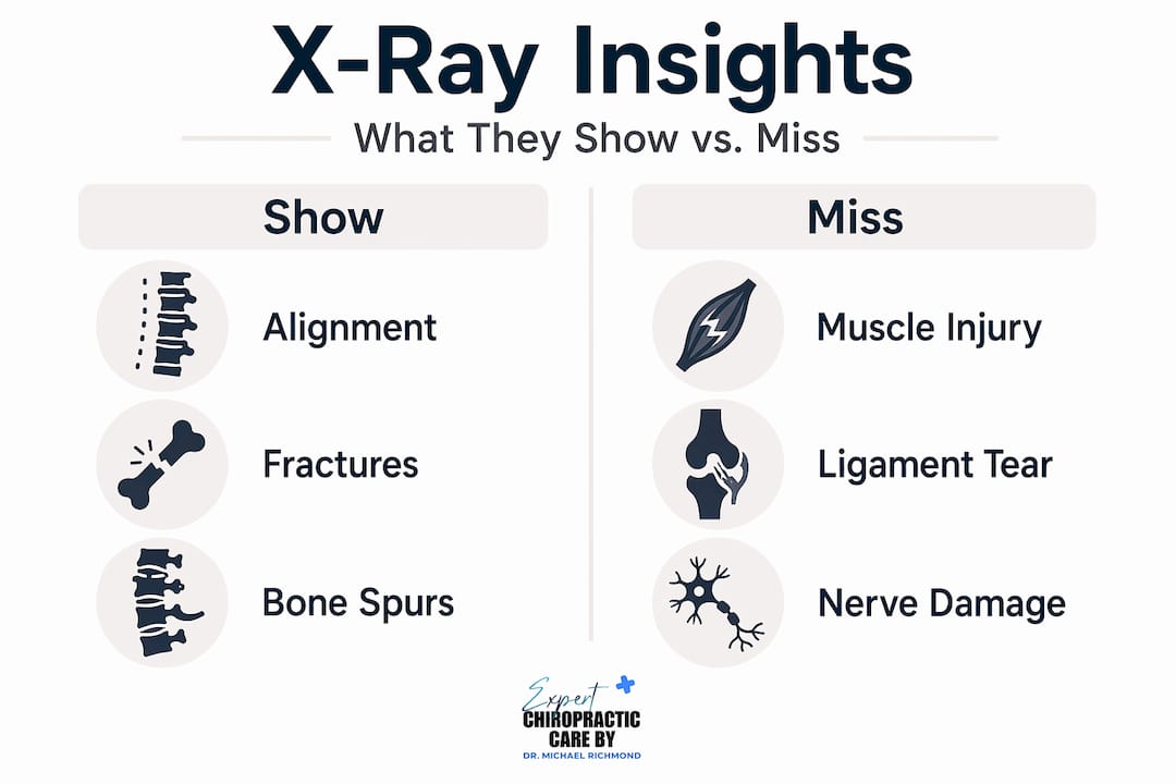
Here is a quick comparison of what standard x-rays show versus what they miss:
| What x-rays reveal | What x-rays cannot show |
|---|---|
| Bone structure and alignment | Soft tissue injuries (muscles, ligaments, tendons) |
| Fractures and dislocations | Disc herniation detail (MRI is preferred) |
| Signs of arthritis or degeneration | Nerve compression specifics |
| Bone tumors or lesions | Early-stage inflammation |

Beyond static images, digital motion x-ray technology is changing what chiropractors can assess. This tool records the spine while you actively flex and extend, capturing movement patterns rather than a frozen snapshot. More on that technology in a later section.
When x-rays are clinically appropriate
Not every chiropractic patient needs imaging. Clinical guidelines advise reserving x-rays for patients who present specific red flags or meet defined criteria. Ordering an x-ray without a clinical reason is not standard practice, and for good reason.
The situations that typically justify imaging in a chiropractic setting include:
- History of cancer or malignancy: Any patient with a prior cancer diagnosis warrants imaging to rule out metastatic bone involvement before spinal manipulation begins.
- Acute trauma: If you were in a car accident, suffered a fall, or experienced any significant impact, fracture screening is a priority before any adjustments.
- Neurological symptoms: Numbness, weakness, or loss of bladder or bowel control alongside back pain signals potential spinal cord involvement that must be investigated.
- Unexplained weight loss or fever combined with back pain: These systemic symptoms could point to infection or malignancy rather than mechanical back pain.
- Failure of conservative care after 4 to 6 weeks: If your pain has not improved after an initial course of chiropractic treatment, imaging helps identify whether a structural reason is at play.
Clinical decision tools like the Ottawa Spine Rules were originally developed for emergency settings, but they have informed how chiropractors think about when imaging crosses from unnecessary to warranted. Insurance and regulatory factors also play a role. Medicare policies require documented clinical justification to authorize imaging, which creates a built-in check against routine overuse.
Pro Tip: Before your appointment, write down any red flag symptoms you have, including recent trauma, unexplained weight loss, or neurological changes. This helps your chiropractor make a faster and more accurate imaging decision.
The Choosing Wisely initiative has been particularly clear: routine full-spine x-rays for nonspecific low back pain without red flags are not backed by evidence. You have every right to ask your chiropractor why imaging is being recommended and what specific finding they are looking for.
Risks and limitations you should know
X-rays are valuable tools. They are also a source of ionizing radiation, which accumulates in your body over a lifetime of exposures. Medical imaging contributes roughly 48% of total population radiation exposure in the United States. That number matters because most radiation exposure happens in small, incremental doses across dozens of medical visits over years.
"The goal is never to avoid imaging when it is clinically needed. The goal is to avoid imaging when it is not." This distinction separates responsible chiropractic practice from outdated routines.
There are a few genuine concerns worth knowing about:
False positives and incidental findings. An x-ray might reveal a minor structural variation, like a small disc space change or a bone spur, that has nothing to do with your current pain. These findings can trigger follow-up tests, additional appointments, and unnecessary anxiety, even when the original pain was completely unrelated.
Psychological impact. Research in musculoskeletal medicine shows that seeing an image of spinal degeneration, even age-appropriate changes, can make patients feel worse and become more fearful of movement. This can slow recovery rather than accelerate it.
Overuse in low-risk patients. For straightforward, uncomplicated low back pain without any red flags, the guidelines are consistent. Imaging adds cost and radiation without improving outcomes. The Choosing Wisely campaign exists precisely because routine x-ray ordering became too common in settings where it added little clinical value.
State radiation safety regulations and professional licensing boards set compliance requirements for x-ray equipment calibration and proper shielding to manage these risks in clinical settings.
How x-ray findings shape your treatment plan
This is where the importance of x-rays in chiropractic care becomes most concrete. An x-ray is not just a diagnostic checkbox. It directly determines what your chiropractor does and does not do during your session.
Consider these scenarios:
- Spondylolisthesis detected: A condition where one vertebra has slipped forward over another. Without that image, a high-velocity adjustment to that area could cause harm. With it, your chiropractor can apply gentler, targeted techniques or refer you to a specialist.
- Severe osteoporosis confirmed: Significant bone density loss rules out traditional spinal manipulation entirely. The treatment shifts toward soft tissue work, low-force techniques, and lifestyle recommendations.
- Post-accident structural changes: For car accident recovery specifically, x-rays help identify whiplash-related cervical misalignments that guide the direction and force of every cervical adjustment. You can learn more about how chiropractic methods post-accident are tailored around imaging findings.
- Scoliosis monitoring: Adolescent scoliosis patients may receive follow-up x-rays at specific intervals to track curve progression, which determines whether conservative chiropractic care remains appropriate or if surgical consultation is needed.
Pro Tip: Ask your chiropractor to show you your x-ray images and explain what they see. Understanding your own structural findings helps you engage with your treatment rather than just receive it.
Follow-up imaging also plays a role in tracking progress. If your chiropractor suspects a structural condition is improving, a comparative x-ray months later can confirm changes objectively, which is far more reliable than pain ratings alone.
Digital motion x-rays and the future of imaging
Standard x-rays capture a single frozen moment. For certain spinal problems, that is not enough. Digital motion x-ray (DMX) technology records your spine as you move, producing a video-like image of how your vertebrae behave during flexion, extension, and rotation.
DMX captures spinal instabilities that are completely invisible on a static image. A vertebra that looks perfectly positioned at rest might shift abnormally during movement, creating nerve irritation and pain that a traditional x-ray would never reveal. This is particularly useful in cases involving:
- Post-traumatic instability after car accidents
- Chronic neck pain with unclear structural causes
- Cases where standard imaging is normal but symptoms persist
- Functional assessment of the cervical spine after whiplash injuries
DMX technology availability is expanding in 2026, with more chiropractic clinics incorporating it as a diagnostic standard rather than a specialty option. The value is not just in identifying problems. It is in documenting them objectively, which matters for insurance claims, legal cases following accidents, and tracking treatment effectiveness over time.
The broader trajectory of chiropractic imaging is toward precision and lower radiation. Ultrasound for soft tissue assessment, cone-beam CT for detailed spinal views, and AI-assisted image analysis are all moving from specialty clinics into general chiropractic practice. You can read more about diagnostic tests in chiropractic care to see how these tools fit together.
My take on x-rays in chiropractic practice
I've seen a lot of patients come in convinced they need a full-spine x-ray before anyone touches their back. I've also seen patients completely surprised when imaging is actually recommended. Both reactions come from the same place: not knowing what imaging is actually for.
In my experience, the true value of chiropractic x-rays is not routine screening. It is targeted diagnosis when something specific is suspected. X-rays help rule out dangerous conditions and confirm structural findings that would otherwise be invisible, which changes the treatment approach in meaningful ways.
What I've found is that patient education changes the conversation entirely. When you understand what your chiropractor is looking for and why, you become a more active participant in your own care. You ask better questions. You push back on unnecessary procedures. You understand why a gentle technique is being used instead of a firm adjustment.
Technology will keep improving. But no image replaces a skilled clinician who listens to your history, performs a thorough physical exam, and decides whether imaging adds real clinical value for your specific situation. X-rays are a tool. How they are used depends entirely on the practitioner wielding them.
— Spark
Ready to learn more about your chiropractic options?
Knowing how imaging works is one part of getting better care. The other part is working with practitioners who use that information thoughtfully.

At Sparkmed, the team in North Miami applies evidence-based diagnostic practices to every patient, whether you are recovering from a car accident, managing chronic back pain, or exploring chiropractic care for the first time. Every treatment plan begins with a thorough clinical assessment, and imaging is recommended only when it genuinely changes how care is delivered. If you are curious about faster recovery through chiropractic treatment or want to explore the full range of diagnostic and treatment tools available, Sparkmed's educational resources are a strong place to start. Book your first visit today, no insurance required.
FAQ
Do chiropractors always take x-rays?
No. Chiropractors only recommend x-rays when specific clinical red flags are present, such as trauma, neurological symptoms, or failure of initial care after 4 to 6 weeks. Routine imaging without a clinical indication is not supported by current guidelines.
What do chiropractors look for on x-rays?
Chiropractors use x-rays to assess spinal alignment, detect fractures, evaluate bone density, and identify structural abnormalities that would influence how or whether spinal manipulation is performed.
Are x-rays safe at a chiropractic office?
Yes, when used appropriately. Chiropractic facilities must meet state radiation safety regulations, and clinical guidelines help minimize unnecessary exposure since medical imaging accounts for a significant portion of cumulative population radiation.
What is a digital motion x-ray?
A digital motion x-ray is an advanced imaging tool that captures the spine in motion rather than in a static position, revealing spinal instabilities and movement dysfunctions that standard x-rays cannot detect.
Can x-ray findings change my chiropractic treatment?
Absolutely. Findings like severe osteoporosis or spondylolisthesis directly modify which techniques a chiropractor uses, often replacing high-force adjustments with gentler, lower-risk alternatives to protect your safety.